
Anatomy of the Chest Muscles in Yogveda Yoga: Pectoralis, Intercostals, and Serratus
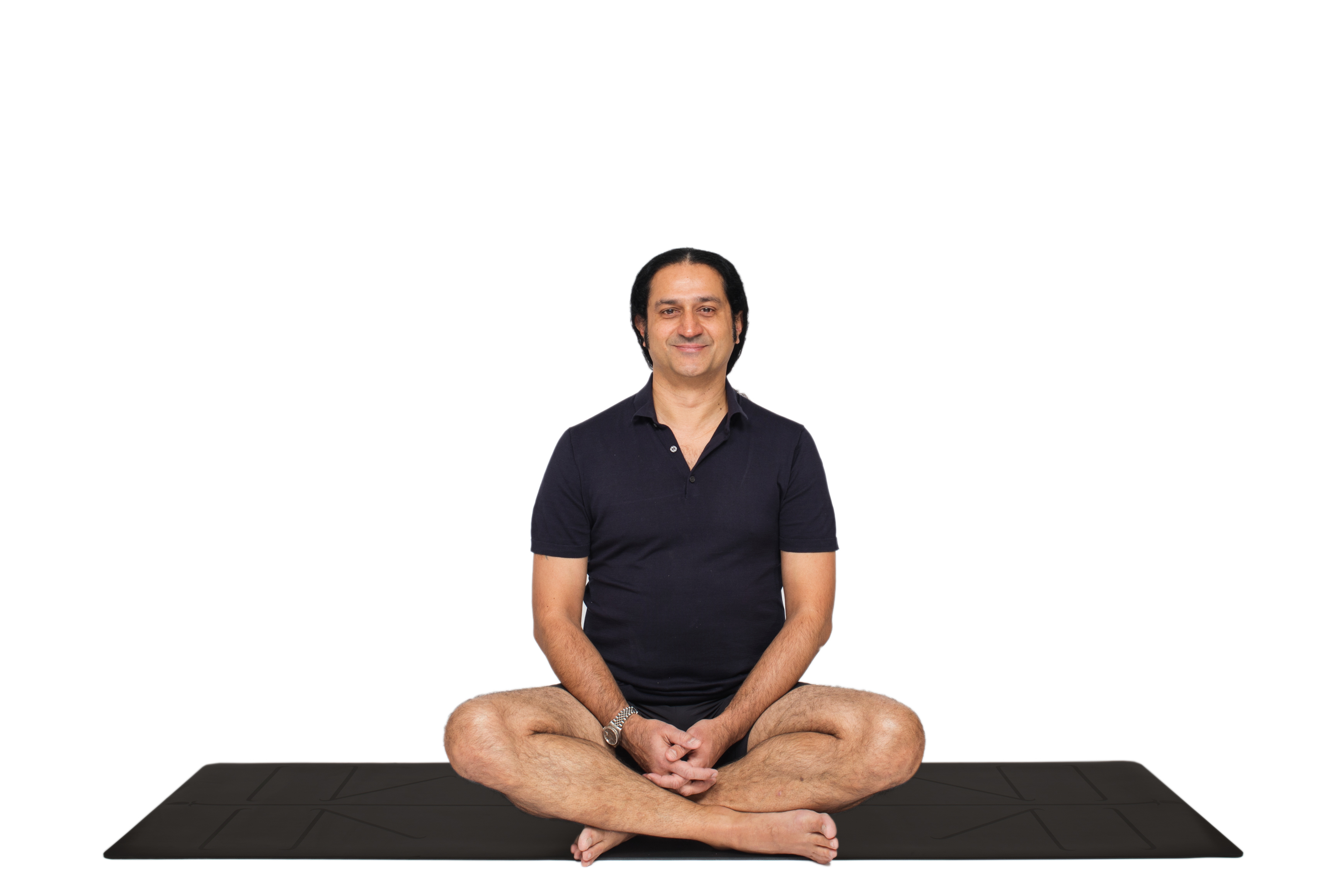

The thoracic cage is a dynamic muscular vault responsible for our breathing mechanics, the regulation of our autonomic nervous system, and the stability of our upper body. In applied biomechanical yoga science, understanding the precise anatomy of the front of the chest is the fundamental first step toward correcting compromised posture and restoring the full natural capacity of our lungs.
To practice the classical science of Patanjali is to understand and command the biological structures that house the breath. The chest musculature is made up of distinct groups that dictate how effortlessly we inhale, how deeply we can exhale, and ultimately, the neurological state of our entire physiology.
This article is the first in a comprehensive five-part series detailing the biomechanics of the thoracic muscles. Over the next four articles, we will take a deep dive into each primary muscle group, examining their specific anatomical functions, how they contribute to respiratory restriction, and their precise application within Yogveda Yoga.
The Four Primary Chest Muscles for Respiratory Mechanics
Pectoralis Minor: The Deep Chest Muscle Restricting Diaphragmatic Breathing
Lying hidden beneath the pectoralis major is the pectoralis minor, a small, triangular muscle that connects the shoulder blade (scapula) to the third, fourth, and fifth ribs. In structural biomechanics, a short and tight pectoralis minor is the silent root cause of severe postural decline. It physically tilts the shoulder blade forward and down, which compresses the chest cavity, collapses the sternum, and severely limits the upward and outward movement of the ribs. If this deep muscle is closed and contracted, it forces the larger superficial chest muscles into a collapsed state. Releasing the pectoralis minor is absolutely critical for restoring structural space for the diaphragm to function.
Pectoralis Major: Anatomy and Function in Postural Alignment
The pectoralis major is the largest and most superficial muscle of the chest wall. Originating from the collarbone, sternum, and upper ribs, it attaches to the upper arm bone (humerus). While its primary anatomical job is to bring the arm across the body and rotate it inward, its biomechanical significance in yoga lies in its resting tension. Because it sits on top of the pectoralis minor, any underlying collapse forces the pectoralis major into a chronically shortened state. This tension acts like a tight band across the chest, locking the shoulders forward and restricting the natural expansion of the rib cage required for deep diaphragmatic breathing.
Intercostal Muscles: The Essential Muscles for Rib Cage Expansion
Situated entirely in the spaces between the ribs, the external and internal intercostal muscles act as the architects of breathing. The external intercostals elevate the ribs during inhalation, actively expanding the width and depth of the chest cavity. Conversely, the internal intercostals draw the ribs downward and inward during forced exhalation. This downward contraction is a mechanism we deliberately engage during specific Pranayama techniques and the prolonged exhalations necessary for calming the nervous system.
Serratus Anterior: Scapular Stabilization for the Thoracic Wall
Spanning the side of the rib cage from the upper eight ribs to the inner edge of the shoulder blade, the serratus anterior anchors the scapula to the back of the chest wall. When this muscle becomes weak, the shoulder blade lifts away from the ribs—a condition known as scapular winging. This lack of stability disrupts the entire upper body, forcing the chest and neck muscles to overwork just to keep the torso upright, which further restricts our breathing efficiency.
Biomechanics of the Chest Muscles and Autonomic Nervous System Regulation
These muscles do not operate in isolation; they function as a continuous, interconnected system. When the chest muscles are chronically tight due to a collapsed posture—such as a rounded upper back (thoracic kyphosis)—our breathing mechanics are severely compromised. The body is forced to rely on smaller accessory muscles in the neck to literally lift the weight of the rib cage with every breath.
This shallow, upper-chest breathing pattern acts as a biological alarm system. It signals a state of physiological threat to the autonomic nervous system, locking the body into sympathetic dominance (fight-or-flight). Applied biomechanical yoga disciplines, particularly Kriya Yoga and classical Ashtanga, utilize highly specific postures and breath control to elongate the pectoralis groups and strengthen the serratus anterior. This anatomical restoration expands the chest vault, allowing the diaphragm to descend fully, which stimulates the vagus nerve and shifts the nervous system back into a state of deep, restorative parasympathetic regulation.
Scientific Evidence for Thoracic Posture and Respiratory Muscle Function
Peer-reviewed research confirms the direct relationship between chest posture, respiratory muscle function, and autonomic regulation. Altered body positions, particularly slouched or collapsed upper body postures, change the resting length of respiratory muscles, making them fundamentally weaker. Studies utilizing Sniff Nasal Inspiratory Pressure (SNIP) measurements demonstrate that compromised sitting postures significantly reduce diaphragmatic tension and overall respiratory muscle strength, leading to a decreased respiratory rate and depth. Furthermore, controlled breathing interventions targeting chest expansion and diaphragmatic engagement have been shown to directly modulate the autonomic nervous system, enhancing heart rate variability and restoring cardiovascular balance.
Citations:
Albarrati, A. M., Zafar, H., Alghadir, A. H., & Anwer, S. (2018). Effect of Upright and Slouched Sitting Postures on the Respiratory Muscle Strength in Healthy Young Males. Annals of Thoracic Medicine, 13(1), 21–25.
Kim, J., et al. (2024). Analysis of breathing patterns to stabilize cardiovascular changes and autonomic nervous system reactivity. Biomedical Signal Processing and Control, 76, 103714.
Yogveda Manifesto "Yogveda Yoga is a compiled applied science. I do not follow modern 'isms,' dogmas, or blind beliefs. Rooted strictly in the classical Darśanas and Ayurveda, and validated by peer-reviewed physiological studies, I dictate no human morality and teach no god worship. I simply provide the exact tools to study your own biological engine. Freedom is absolute self-knowledge. Study yourself to be free." — Master Shahid Khan
Yogveda Yoga Switzerland 🇨🇭 To establish an official Yogveda Yoga School in your city or country, please direct your inquiry to info@yogveda.ch.
Author: Master Shahid Khan Official Portals:

Comments